
Biomedical Scientific Illustration

22 archived cases

Compare the visible organization of research objects, processes, interfaces and local views. Each detail page retains its original artwork and associated paper record.

Discuss a paper figure
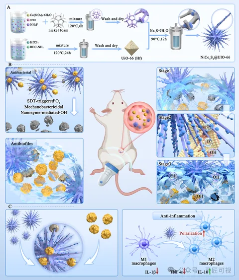
Piezoelectric Nano-Urchins for Drug-Resistant Biofilm Infections

Piezoelectric Nano-Urchins for Drug-Resistant Biofilm Infections

Material preparation runs horizontally across the top. An animal application connects to the central scene, with functional regions on the left and stage panels on the right. Read preparation and application location before the local stages and lower results.

Biomedical researchScientific paper figurePublished work
Three Routes for Extracting RG-I-Enriched Apple Pectin

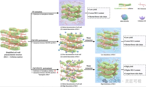
Three Routes for Extracting RG-I-Enriched Apple Pectin

A shared cell-wall structure on the left branches into three treatment routes. Resulting structures and result boxes occupy the right. The common starting point, aligned stages and consistent color roles support comparison.

Biomedical researchScientific paper figurePublished work
Mitochondria-Targeted Tannic Acid–Strontium Nanomedicine

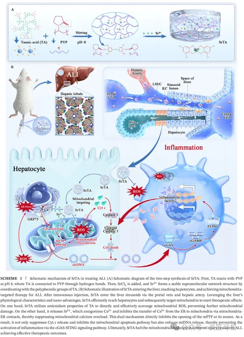
Mitochondria-Targeted Tannic Acid–Strontium Nanomedicine

Material preparation sits above an animal-to-liver view. Tissue details lead into cellular and organelle panels below. Insets and arrows separate the material, application location and intracellular relationships across scales.

Biomedical researchScientific paper figurePublished work
Recombinant Collagen Hydrogel and Infected Wound Repair

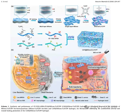
Recombinant Collagen Hydrogel and Infected Wound Repair

A horizontal preparation workflow and converging components introduce the composite above. Bacterial detail and a wound scene sit together below, connected through frames, arrows and local insets with a shared legend.

Biomedical researchScientific paper figurePublished work
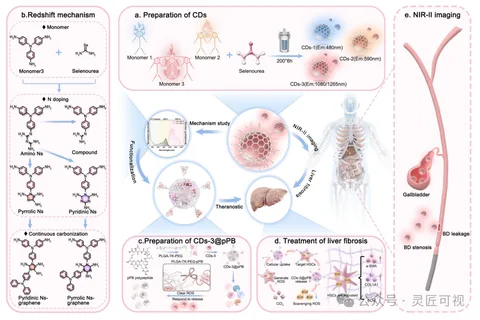
NIR-II Carbon Dots for Hepatobiliary Theranostics

NIR-II Carbon Dots for Hepatobiliary Theranostics

Lettered panels separate carbon-dot preparation, structural mechanisms and an imaging scene. A central object connects applications, while functionalization and local application information continue along the bottom.

Biomedical researchScientific paper figurePublished work
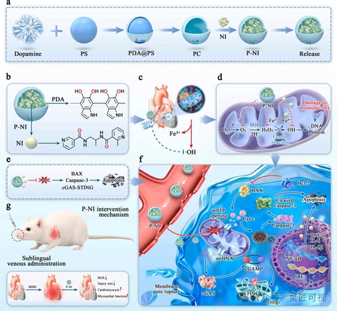
ROS-Scavenging Nanocarriers and Myocardial Injury

ROS-Scavenging Nanocarriers and Myocardial Injury

Sequential steps across the top show nanocarrier preparation. Lettered regions connect the material structure, heart location and intracellular information. A larger cellular inset holds the dense relationships, with smaller panels for administration and results.

Biomedical researchScientific paper figurePublished work
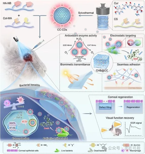
Enzyme-Mimetic Hydrogel for Keratitis and Corneal Regeneration

Enzyme-Mimetic Hydrogel for Keratitis and Corneal Regeneration

Material components above lead into an eye application. Several insets show local material and cellular information, with lines, arrows and dashed frames connecting levels. A separate results illustration sits at the lower right.

Biomedical researchScientific paper figurePublished work
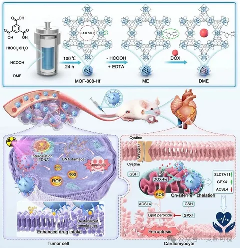
EDTA-Functionalized MOFs for Tumor Therapy and Cardiac Protection

EDTA-Functionalized MOFs for Tumor Therapy and Cardiac Protection

MOF structural changes occupy the top. The middle links animal, tissue and heart views, followed by two cellular environments side by side below. Background colors and outlines distinguish each region.

Biomedical researchScientific paper figurePublished work
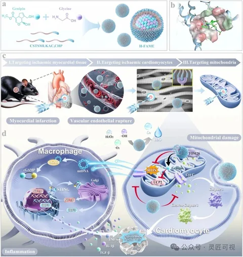
Heart-Targeted Nanomedicine and Mitochondrial Damage

Heart-Targeted Nanomedicine and Mitochondrial Damage

Assembly and structural details appear above a sequence linking the heart, cells and mitochondria. Two cellular environments follow below. Stage bars, insets and arrows establish an overview-to-detail path.

Biomedical researchScientific paper figurePublished work
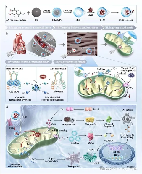
mitoNEET-Related Nanotherapeutics for Myocardial Injury

mitoNEET-Related Nanotherapeutics for Myocardial Injury

The top sequence establishes carrier assembly before stage bars and insets lead to cells and mitochondria. A protein-structure comparison and a larger cellular mechanism panel hold separate details, coordinated by legends, arrows and numbering.

Biomedical researchScientific paper figurePublished work
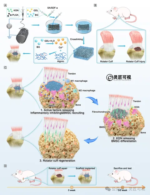
Hydrogel–Nanofiber Scaffolds for Rotator Cuff Repair

Hydrogel–Nanofiber Scaffolds for Rotator Cuff Repair

Separate preparations for a fibrous membrane and hydrogel lead to the rotator-cuff injury location. Three local stages organize tissue changes, while a lower timeline records implantation and observation arrangements.

Biomedical researchScientific paper figurePublished work
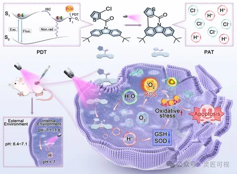
Organic Photosensitizers: ROS and Proton Co-Generation

Organic Photosensitizers: ROS and Proton Co-Generation

Small upper blocks hold energy levels, molecular structures and ionic information. An illuminated animal scene connects the application to a larger lower-right cell, where ROS, protons and arrows explain local relationships.

Biomedical researchScientific paper figurePublished work
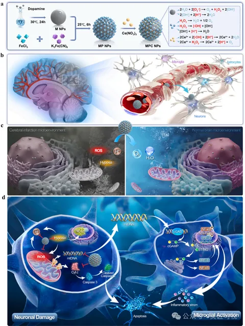
Sequentially Targeted Nanomedicine and the Neural Microenvironment

Sequentially Targeted Nanomedicine and the Neural Microenvironment

A top preparation workflow leads to brain and vessel locations. Two local environments are compared in the middle. Larger neuron and microglial panels below hold the molecular pathways.

Biomedical researchScientific paper figurePublished work
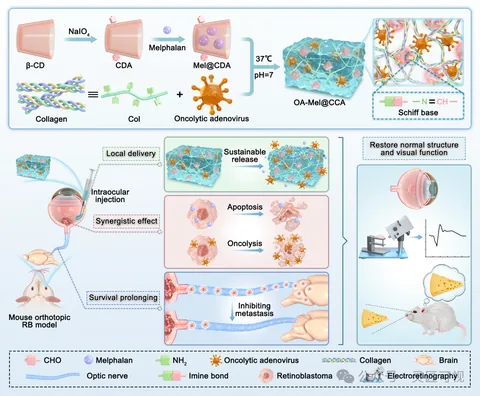
Hydrogel Co-Delivery of Oncolytic Adenovirus and Melphalan

Hydrogel Co-Delivery of Oncolytic Adenovirus and Melphalan

Two component paths converge into a gel network above. An animal and eye locate the injection below, followed by separate release and cellular regions. A right-hand eye and assessment scene share the object legend.

Biomedical researchScientific paper figurePublished work
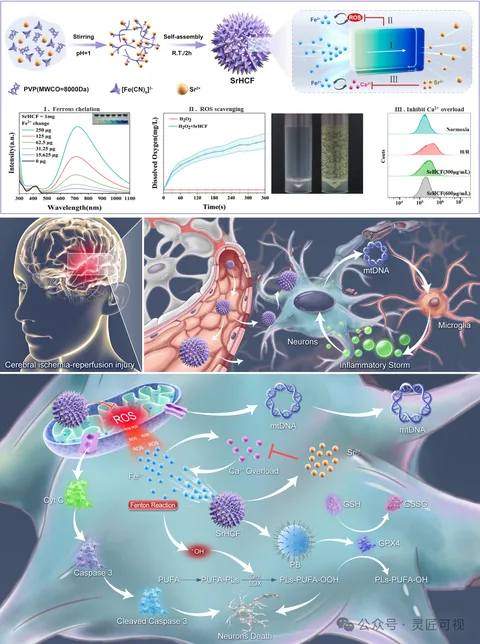
Strontium-Substituted Nanomedicine for Cerebral Injury

Strontium-Substituted Nanomedicine for Cerebral Injury

Particle assembly and characterization sit at the top. Brain injury, vessel and neuron views lead to a large lower cellular panel. Curves, apparatus and mechanism arrows occupy separate regions.

Biomedical researchScientific paper figurePublished work
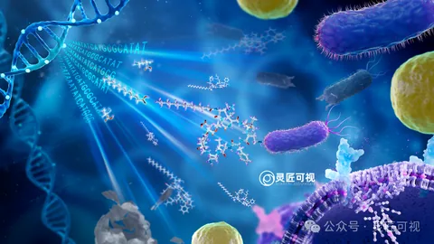
Dual-Target Antibiotic: Molecules, Bacteria and Membranes

Dual-Target Antibiotic: Molecules, Bacteria and Membranes

DNA and sequence text in the upper left introduce a molecular structure. Bacteria of different sizes extend to the right and distance, while a membrane below supplies a local scale. Left-to-right light connects the scene.

Biomedical researchScientific paper figurePublished work
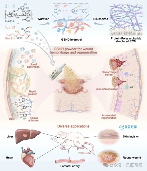
Rapid-Gelling Hyaluronan Hemostatic Powder

Rapid-Gelling Hyaluronan Hemostatic Powder

Two components lead into a hydrogel and network above. The central wound connects absorption and adhesion on the left with two local stages on the right. Smaller organ and wound views show application locations below.

Biomedical researchScientific paper figurePublished work
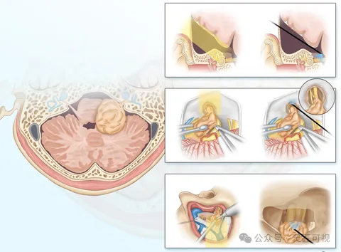
Neuroendoscopy in Vestibular Schwannoma Resection

Neuroendoscopy in Vestibular Schwannoma Resection

Both illustrations use a large intracranial cross-section for orientation. Grouped panels on the right show instruments, views and local anatomy. Corresponding perspectives, letters and original captions support regional comparison.

Biomedical researchScientific paper figurePublished work
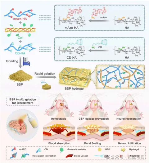
Self-Gelling Adhesive Powder for Brain-Injury Sealing

Self-Gelling Adhesive Powder for Brain-Injury Sealing

Three components form powder and gel above, with a network inset on the right. The lower half moves from a brain location to three local scenes for hemostasis, sealing and neural information. A shared legend links the levels.

Biomedical researchScientific paper figurePublished work
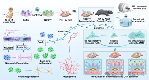
Sox2-Overexpressing Neural Stem Cells and Hydrocephalus

Sox2-Overexpressing Neural Stem Cells and Hydrocephalus

Upper arrows connect cell source, treatment, model and transplantation before imaging and behavioral assessments. Lower regions separate signaling, neural, immune and local-tissue information with dashed borders.

Biomedical researchScientific paper figurePublished work
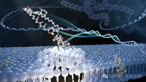
Mandimycin and Fungal Membrane Phospholipids

Mandimycin and Fungal Membrane Phospholipids

A foreground ball-and-stick molecule extends toward a membrane, where selected phospholipids are enlarged. Lower-contrast fungal chains and a double helix remain in the background.

Biomedical researchScientific paper figurePublished work
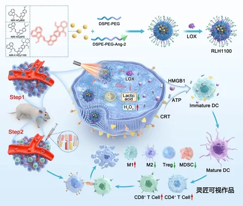
Hemicyanine Nanoparticles and Glioblastoma Photoimmunotherapy

Hemicyanine Nanoparticles and Glioblastoma Photoimmunotherapy

An upper assembly sequence connects to two labeled delivery stages and a vessel. A larger cell on the right holds the main relationships; different immune-cell forms and arrows continue below.

Biomedical researchScientific paper figurePublished work