
Materials Chemistry Visualization

7 archived cases

Compare the visible organization of research objects, processes, interfaces and local views. Each detail page retains its original artwork and associated paper record.

Discuss a paper figure
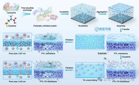
Amyloid-Like Assembly of Protein Nanofiltration Membranes

Amyloid-Like Assembly of Protein Nanofiltration Membranes

Protein structures at the top lead through nucleation and assembly, then transfer and crosslinking to membranes below. Paired membrane surfaces and pore details distinguish process order from structural comparison.

Materials chemistryScientific paper figurePublished work
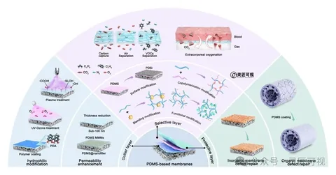
PDMS Gas-Separation Membranes: A Semicircular Review Figure

PDMS Gas-Separation Membranes: A Semicircular Review Figure

The PDMS base membrane and layer roles occupy the lower center. Outer sectors organize hydrophilic modification, permeability, copolymerization and defect repair. Gas-separation applications sit above, creating an inside-to-outside hierarchy.

Materials chemistryScientific paper figurePublished work
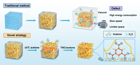
Ice-Dissolving Crosslinking of Polyelectrolyte Monoliths

Ice-Dissolving Crosslinking of Polyelectrolyte Monoliths

Two rows begin with similarly viewed porous blocks. The upper route connects a vacuum setup and annotation box; the lower route shows solvent treatment and crosslinking. A right-hand inset links macropores to polymer chains and junctions.

Materials chemistryScientific paper figurePublished work
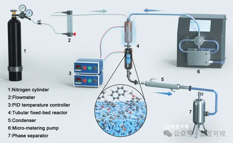
Catalytic Methylation: Reactor Apparatus and Local Structure

Catalytic Methylation: Reactor Apparatus and Local Structure

Gas enters from the left through pipes around a central fixed-bed reactor. Numbered controllers, condenser, pump and separator provide lookup points. A separate circular inset below shows internal microscopic structure.

Materials chemistryScientific paper figurePublished work
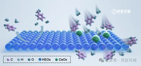
Cerium-Doped High-Entropy Oxides: A Catalytic Interface

Cerium-Doped High-Entropy Oxides: A Catalytic Interface

Blue spheres form a continuous surface, with a smaller number of green spheres marking selected sites. Ball-and-stick molecules sit above the surface. Curved arrows distinguish approach, surface processes and departure, supported by a lower legend.

Materials chemistryScientific paper figurePublished work
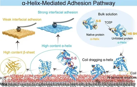
α-Helix-Mediated Protein Adhesion

α-Helix-Mediated Protein Adhesion

The first illustration connects protein structural changes to an interface and compares two adhesion states. The second places surface structures, protein insets and plotted results together, using yellow and blue to link corresponding information.

Materials chemistryScientific paper figurePublished work
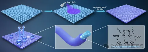
Fluorine-Free Waterborne Fabric Coatings

Fluorine-Free Waterborne Fabric Coatings

Blue fabric progresses through spraying and drying to a purple surface above. Droplets and a cross-section below connect appearance to the coating interface, while a separate right-hand frame holds molecular structures.

Materials chemistryScientific paper figurePublished work