
Bring a documented structure into a scientific figure
Choose the molecule, crystal or protein workflow by the object you need to explain. Record the structure source, choose the view and representation, and check scene editability separately from the final image.
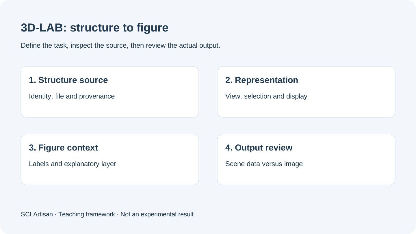
Start with three kinds of structural context
The product distinguishes molecule, crystal and protein workflows. Use the source object to select the workflow, then check which inputs the current interface accepts. The existence of a workflow does not establish universal support for every file format or every representation.
| Object | Record before rendering | Figure review |
|---|---|---|
| Molecule | Identity, source and structural assumptions | Atoms, bonds, labels and view |
| Crystal | Structural record and periodic context | Unit-cell context and selected region |
| Protein | Accession or file source and selection | Chains, residues, representation and view |
When using a database record, retain its identifier and the relevant source version or retrieval information. The RCSB PDB introductory guide explains the context of PDB data; it does not certify any newly added arrows or interpretations in your figure.
Separate a source record from a presentation choice
Keep at least two records: the original structure source and the scene used for presentation. A view direction, color assignment or simplified representation can improve readability without creating new evidence. Record which objects were hidden or emphasized, especially when a selection could affect interpretation.
Do not use a visually plausible distance, bond or interface as proof of a reaction, affinity or therapeutic effect. Those statements need their own scientific basis and author review. The figure caption should make the distinction between a structure display and a conceptual mechanism clear.
Add an explanatory layer around the structural view
A scientific figure often needs three components: the structure view, a readable label layer and an explanation of the setting or relationship. Keep the labels and arrows consistent with the rest of the paper. A change in the apparent scale should not make two objects look directly comparable when they are not.
The catalytic-interface case note explains how a local representation connects to an overall setting. The archived artwork is used as a composition reference. It is not presented as output generated by the online platform, and the note adds no catalytic-performance result.
Preserve the distinction between a LAB object and an image
An object retaining LAB scene information can support returning to the structural editing workflow. A bitmap can preserve only the rendered appearance. Moving an image on a canvas does not restore atoms, bonds, scene settings or an original structure file.
This matters when agreeing a deliverable. “Editable” may refer to the source scene, the arrangement on the figure canvas, labels in an exported document, or all three. State which form is needed before preparing a commissioned mechanism figure.
Check five things in the delivered file
- The structure identity and selection match the retained source.
- The view and representation communicate the intended object.
- Labels and symbols remain legible at the final figure size.
- The scene, project and exported file have a documented relationship.
- The output meets the target journal's current instructions.
Nature's figure specifications are a source for that publisher's requirements, not a universal rule for every journal. Preserve an editable working version and inspect the actual delivery file. The platform page explains why this preview does not yet open an overseas account or guarantee a live export service.
Frequently asked questions
Which 3D-LAB workflow should I use for a molecular or crystal figure?
Choose by the scientific object and source data. A molecule, periodic crystal and protein structure have different context and display needs; visual appearance alone is not enough.
Does a rendered protein structure establish a binding mechanism?
No. A structure view is a presentation of source information. Binding and mechanism claims require the relevant experimental, computational or literature evidence.
Can I edit a 3D-LAB object after placing it on the figure canvas?
Preserved LAB scene data can support returning to the structural workflow. A flattened image is not the same as a scene or structure source; check the actual object type.
Will SVG export make every atom and 3D label separately editable?
Do not infer that from the file extension. Check the chosen display and output mode, then inspect the actual file for embedded images, paths and text.
Sources and scope
Sources apply within their stated scope. Archive records describe existing work; international account access and checkout remain closed.
Choose the next step
Understand the platform · Scope a design commission · Contact support