
Hydrogel Scientific Figure Cases

6 archived cases

Compare the visible organization of research objects, processes, interfaces and local views. Each detail page retains its original artwork and associated paper record.

Discuss a paper figure
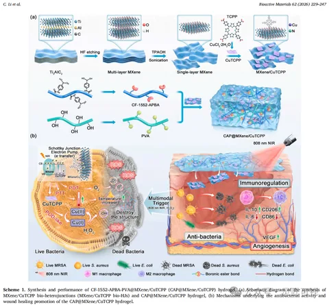
Recombinant Collagen Hydrogel and Infected Wound Repair

Recombinant Collagen Hydrogel and Infected Wound Repair

A horizontal preparation workflow and converging components introduce the composite above. Bacterial detail and a wound scene sit together below, connected through frames, arrows and local insets with a shared legend.

Biomedical researchScientific paper figurePublished work
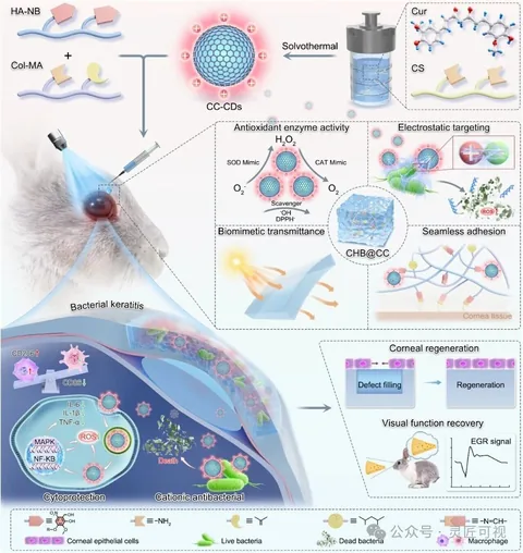
Enzyme-Mimetic Hydrogel for Keratitis and Corneal Regeneration

Enzyme-Mimetic Hydrogel for Keratitis and Corneal Regeneration

Material components above lead into an eye application. Several insets show local material and cellular information, with lines, arrows and dashed frames connecting levels. A separate results illustration sits at the lower right.

Biomedical researchScientific paper figurePublished work
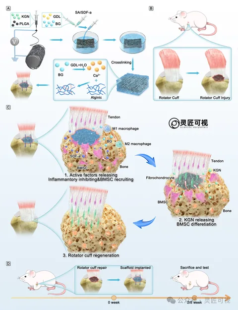
Hydrogel–Nanofiber Scaffolds for Rotator Cuff Repair

Hydrogel–Nanofiber Scaffolds for Rotator Cuff Repair

Separate preparations for a fibrous membrane and hydrogel lead to the rotator-cuff injury location. Three local stages organize tissue changes, while a lower timeline records implantation and observation arrangements.

Biomedical researchScientific paper figurePublished work
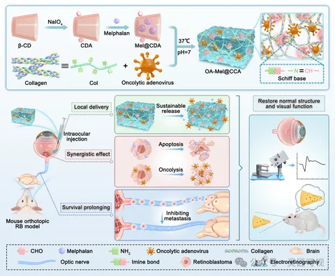
Hydrogel Co-Delivery of Oncolytic Adenovirus and Melphalan

Hydrogel Co-Delivery of Oncolytic Adenovirus and Melphalan

Two component paths converge into a gel network above. An animal and eye locate the injection below, followed by separate release and cellular regions. A right-hand eye and assessment scene share the object legend.

Biomedical researchScientific paper figurePublished work
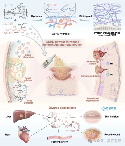
Rapid-Gelling Hyaluronan Hemostatic Powder

Rapid-Gelling Hyaluronan Hemostatic Powder

Two components lead into a hydrogel and network above. The central wound connects absorption and adhesion on the left with two local stages on the right. Smaller organ and wound views show application locations below.

Biomedical researchScientific paper figurePublished work
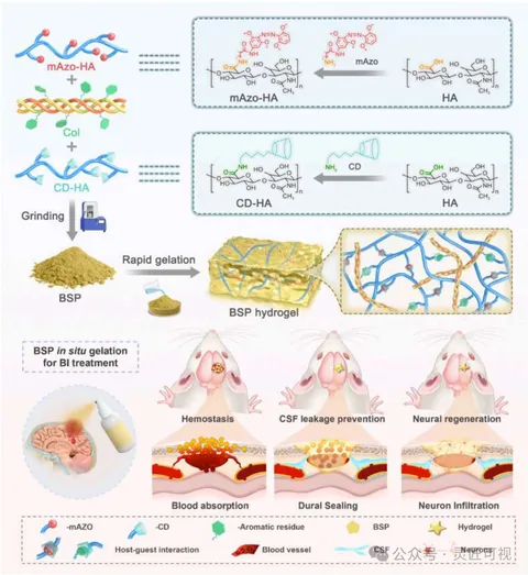
Self-Gelling Adhesive Powder for Brain-Injury Sealing

Self-Gelling Adhesive Powder for Brain-Injury Sealing

Three components form powder and gel above, with a network inset on the right. The lower half moves from a brain location to three local scenes for hemostasis, sealing and neural information. A shared legend links the levels.

Biomedical researchScientific paper figurePublished work