
Review Figure Submission: Specifications, Sources and Attribution
Check the journal’s actual artwork rules, literature support and image permissions separately. A review layout does not establish compliance or permission to reuse its source material.
Record the applicable instructions
Keep the journal, article type, submission stage and current official source together. Identify dimensions, file types, effective resolution, type and color requirements for the specific figure category. General publisher guidance does not replace a journal-specific instruction or an editor's request.
Separate four kinds of source information
Record literature references supporting categories, original data supporting plotted values, licenses for incorporated assets and attribution for existing artwork. These sources serve different purposes. Citing a paper does not automatically grant a right to copy its figure.
Read an archived composition within its scope
The PDMS membrane review figure provides an example of membrane roles, strategies and applications arranged at different levels. Its structure can inform planning; the associated paper and artwork permissions remain separate checks.
Read four examples from published reviews
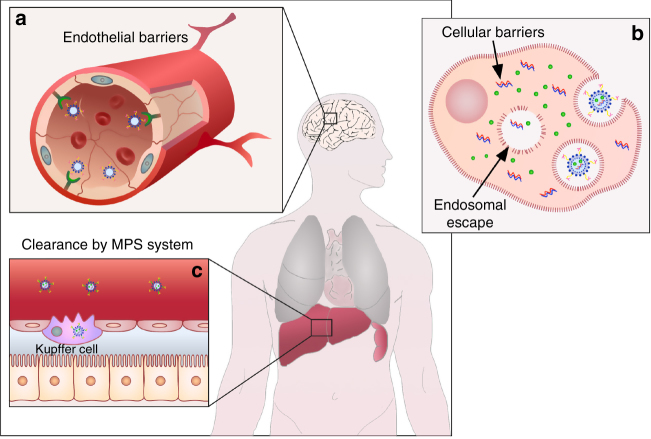
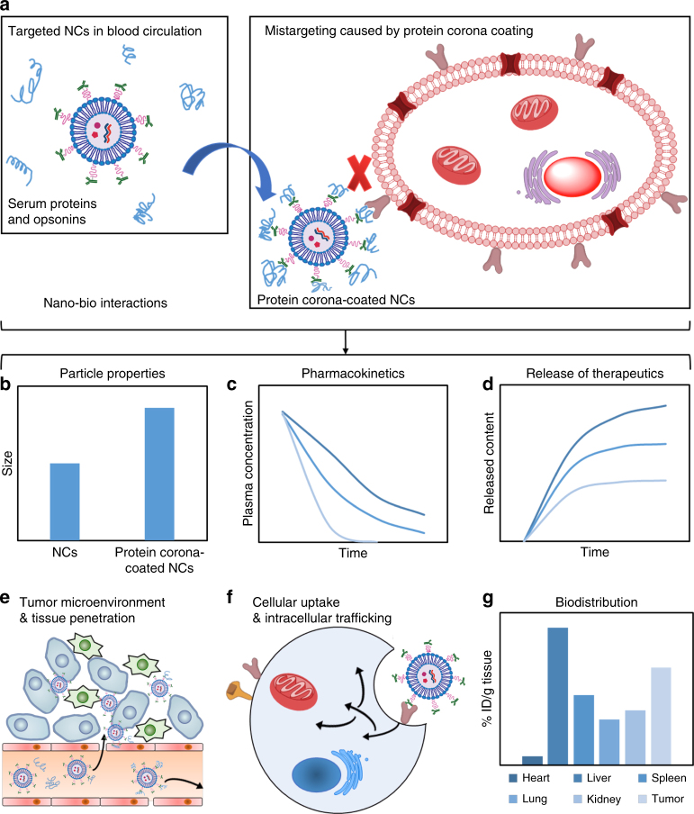
Rosenblum and colleagues’ Nature Communications Figure 1 uses a central body location to organize local delivery barriers. Figure 2 retains a common nanocarrier reference while giving size, pharmacokinetics, release, uptake and distribution their own panels. The shared object supports comparison without implying that every panel answers the same question.
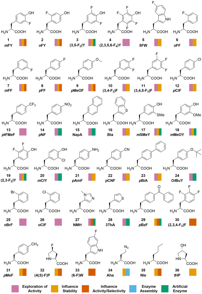
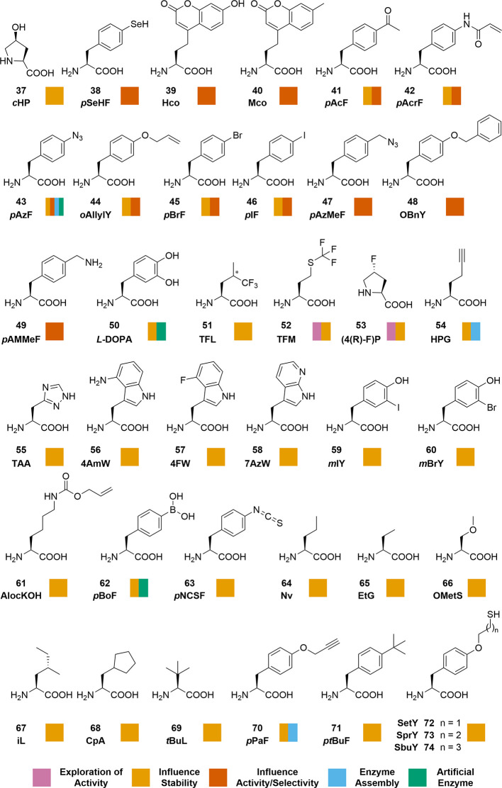
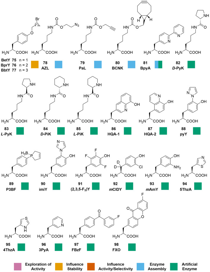
Brouwer and colleagues’ Chemical Reviews Figure 1 uses a numbered structure catalog and color-coded uses. Its three consecutive source image parts remain in their original order below. The layout demonstrates lookup and grouping, rather than a measured proportion of applications.
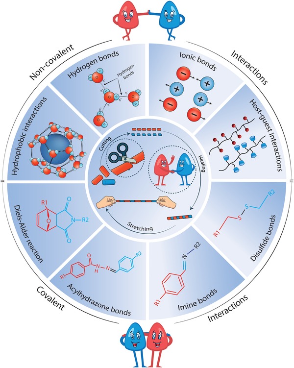
Talebian and colleagues’ Advanced Science Figure 3 places hydrogel cutting, healing and stretching near the center, with interaction and dynamic-bond mechanisms around it. The circular arrangement expresses a common topic and neighboring categories; sector area is not a quantitative result.
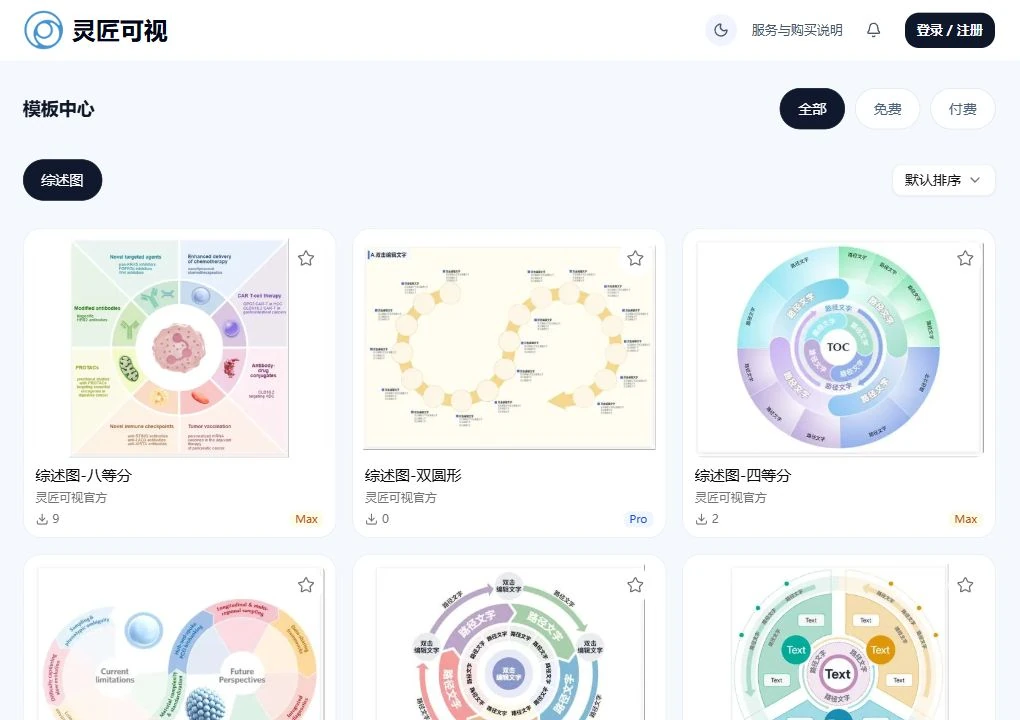
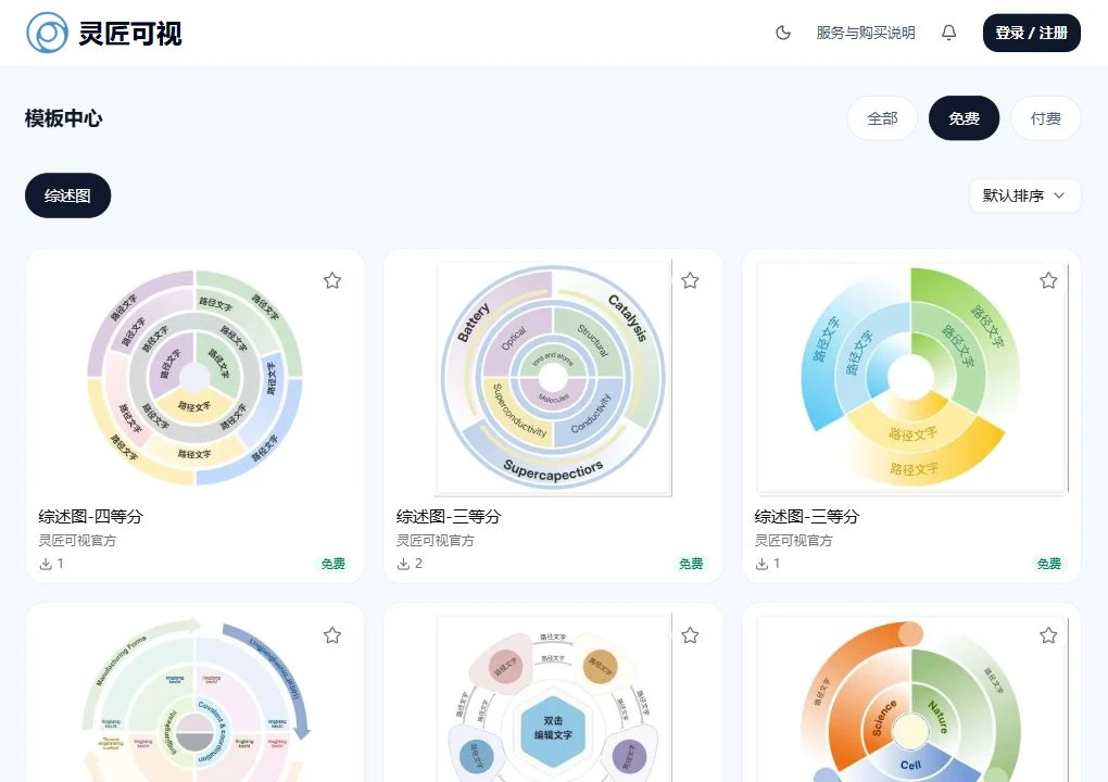
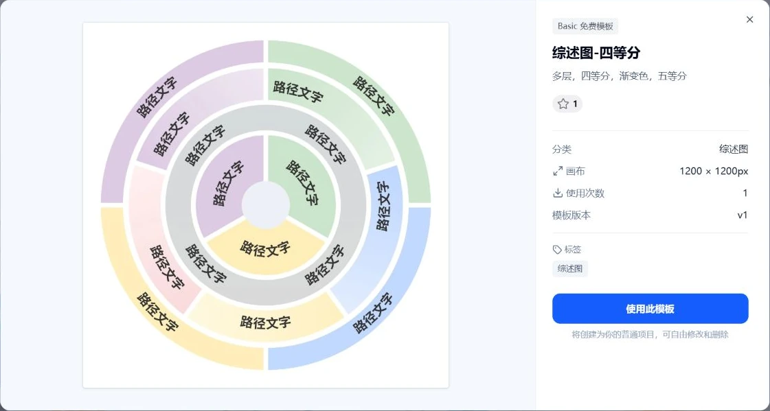
These publisher figures are by their credited authors, not the Lingjiang Keshi team or platform users. Each image retains its own paper link, copyright notice and CC BY 4.0 link. Scientific content has not been redrawn. The China-service template screenshots retain their 16 October 2026 date and are separate from the research examples.
Build a handoff checklist
Record the manuscript version, approved labels, reference mapping, asset permissions, target dimensions, output file and editable source. Reopen the actual export and check font substitution, clipped labels, raster elements and caption references. Ask the corresponding author to approve scientific grouping and any proposed relationships.
Use the journal resource hub, submission checklist and review-figure service scope. The online review workflow explains how to keep a project separate from its export.

Sources and scope
- Nature Reviews artwork guide
- Nature figure specifications
- Nature image-integrity guidance
- Rosenblum et al., targeted delivery review
- Brouwer et al., noncanonical amino acids review
- Talebian et al., self-healing hydrogels review
Sources apply within their stated scope. Archive records describe existing work; international account access and checkout remain closed.
Choose the next step
Understand the platform · Scope a design commission · Contact support