
Multi-Panel Scientific Figures: Layout, Labels and Captions
Give each panel a question, define the reading order and assign space by information need. Coordinate labels and captions before inspecting the figure at its final publication size.
Write one question per panel
List each panel's purpose and source. A preparation route, structural detail, microscopy image and quantitative plot may belong in the same figure, but they should remain identifiable as different evidence or explanation types. Remove duplicate information before allocating space.
Choose the reading order before the grid
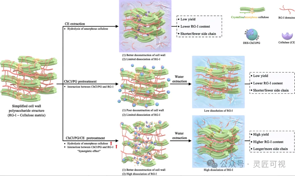
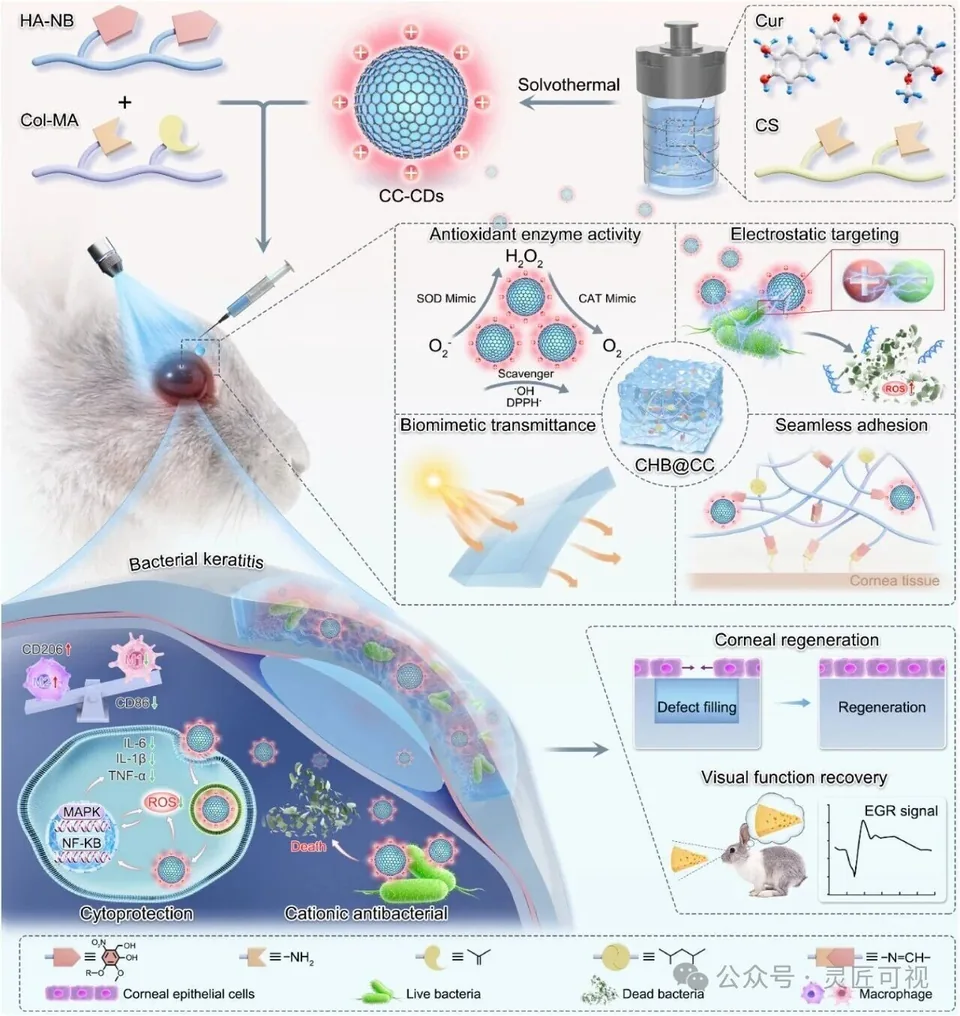
Use left-to-right order for parallel routes when appropriate, or top-to-bottom order for preparation followed by application and local detail. In the apple-pectin case, three routes share one starting reference and aligned result areas. In the hydrogel case, an overview locates several enlargements.
Allocate size by content
Panels need not be equal rectangles. A dense mechanism may need more room than an orientation panel. Preserve comparable scale or explicitly explain differences when comparing images. Keep gutters and alignment consistent without stretching original images to fill a box.
Coordinate numbering and captions
Confirm the panel-letter convention in the journal instructions. Keep letters, abbreviations, units, scale bars and caption references consistent with the manuscript. Distinguish the full-figure caption from explanatory text inside a panel.
Run a final-size inspection
Check text, contrast, object boundaries and the relationship between insets and their source regions. Review original pixels and effective resolution for embedded images. Retain the source file and open the exported deliverable independently. The beginner layout workflow addresses practical arrangement; a TOC submission is a different composition task.
Sources and scope
- Nature figure-panel construction and export
- Nature figure specifications
- Nature image-integrity guidance
Sources apply within their stated scope. Archive records describe existing work; international account access and checkout remain closed.
Choose the next step
Understand the platform · Scope a design commission · Contact support