
Hydrogel Mechanism Figures: Networks, Insets and Release
Separate hydrogel components, network formation, application location and local processes. Use connected insets and defined arrows instead of combining every scale in one scene.
Identify the components and network claim
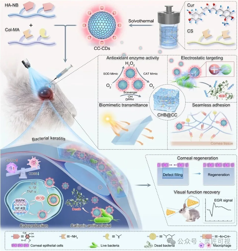
Record polymer or building-block names, relevant bonds or interactions and the evidence supporting the proposed network. A drawn mesh is an explanatory abstraction; its line spacing does not establish measured pore dimensions. Distinguish chemical crosslinks, physical associations and illustrative connections where relevant.
Connect material and application without merging scales
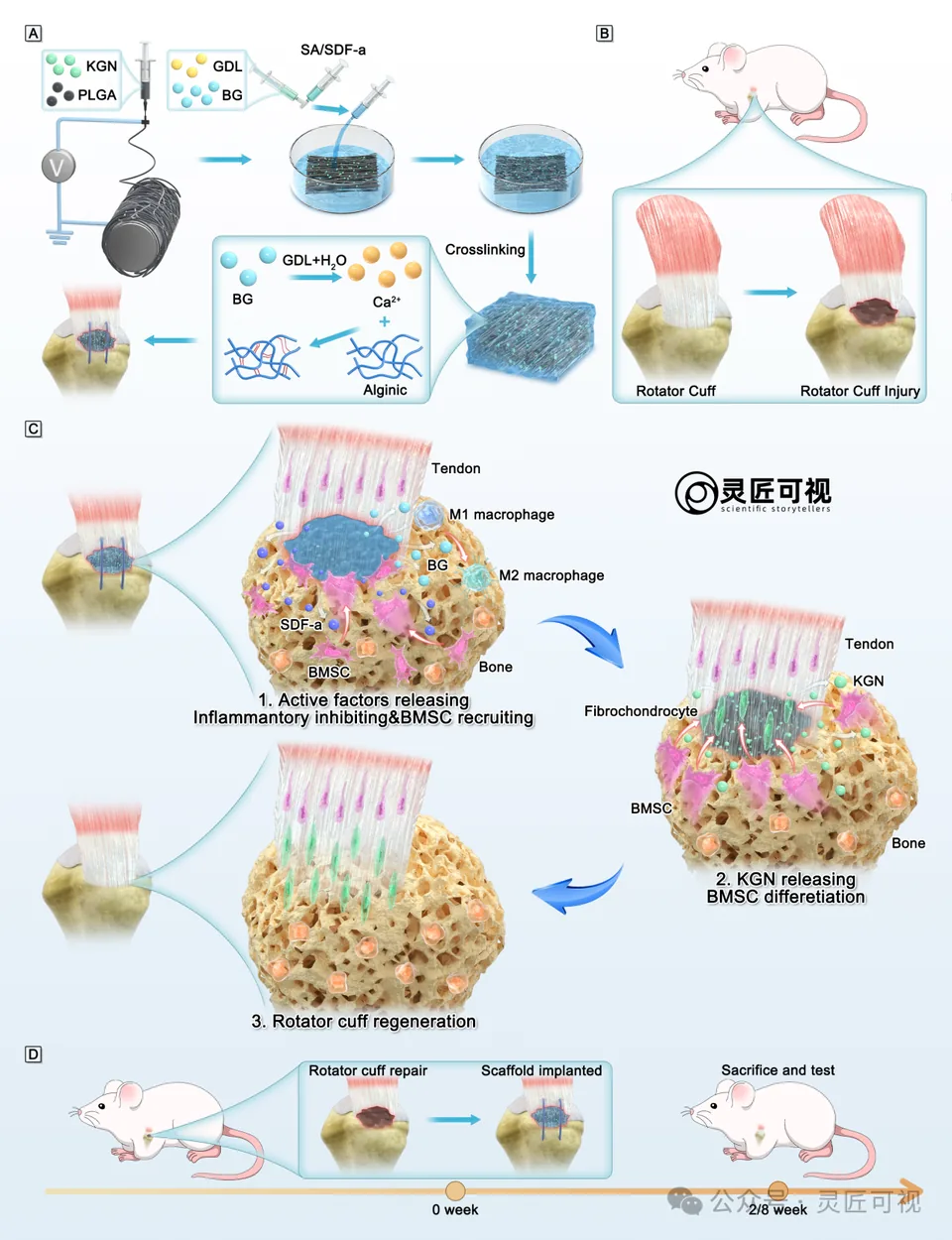
The ACS Nano corneal case places components above an eye application and uses several local views for material and cellular detail. The collagen hydrogel archive connects preparation, composite structure and a wound scene.
Define release and response arrows
A release arrow can indicate transfer of a named component. A cellular arrow may indicate a supported interaction or a proposed explanation. Keep these meanings separate and preserve the source evidence. If the figure compares conditions or time points, use comparable fields of view and unambiguous labels.
Keep the inset tied to its source
Mark which part of the material or tissue is enlarged. Reuse object identity between overview and inset. The image-inset guide explains why display enlargement does not create new image detail or establish biological magnification.
Complete the scientific and file checks
Verify names, network relationships, application location, symbols, permissions and final-size type. Retain a source record for any structure, microscopy or quantitative result. Continue with mechanism planning, panel layout or a reviewed design brief.
Sources and scope
Sources apply within their stated scope. Archive records describe existing work; international account access and checkout remain closed.
Choose the next step
Understand the platform · Scope a design commission · Contact support